Several steps should be performed for diagnosis of lung
cancer:
1.
Careful history taking from the patient by
an experienced physician: the main presenting symptoms include
cough, dyspnea (difficulty breathing), weakness, weight loss,
hemoptysis (coughing up blood), chest pain and hoarseness (due to
involvement of specific nerves adjacent to the tumor).
2.
Careful physical examination should be
performed. Findings on examination may include cachexia (severe body
weight loss), pallor (anemia), tachypnea (increased breath rate),
hoarseness, enlarged lymph nodes, wheezes (due to bronchial
obstruction), pneumonia (due to airways obstruction), and clubbing
of fingers. On auscultation wheezes, or decreased breath sounds (due
to pleural effusion) may be noted.
3.
Laboratory tests may reveal anemia of
malignant disease; leukocyte count can be normal or elevated
(especially if pneumonia is also detected); hyponatremia (low blood
sodium level) is not uncommon, and is mainly due to inappropriate
secretion of anti diuretic hormone (ADH) by tumor cells;
hypercalcemia (elevated blood calcium level); elevated LDH levels.
4.
Radiological evaluation should include
initially chest x-ray and computerized tomography (CT) of the chest.
PET-FDG scan, and fusion of PET-CT scans, can be helpful in detecting metastatic disease.
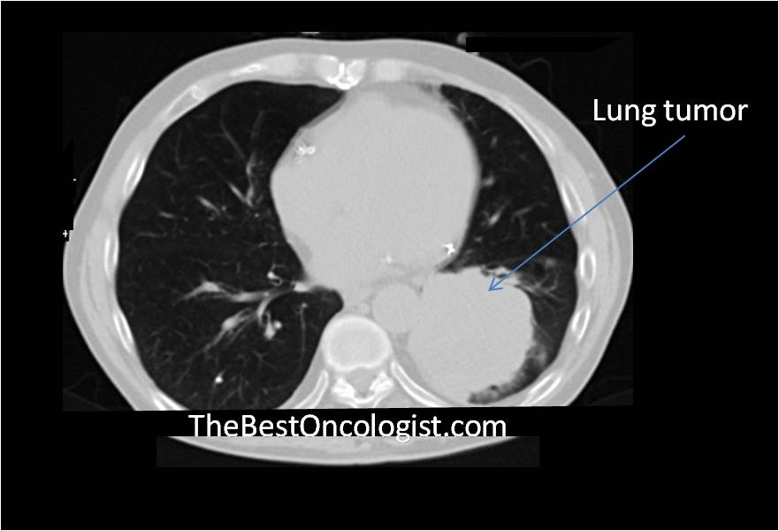
Chest CT
scan showing a lung tumor
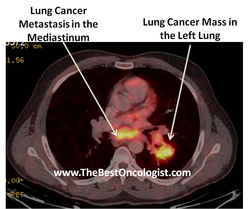
PET-CT Scan: LUNG CANCER TUMOR in the LEFT
LUNG WITH METASTASIS to the MEDIASTINUM
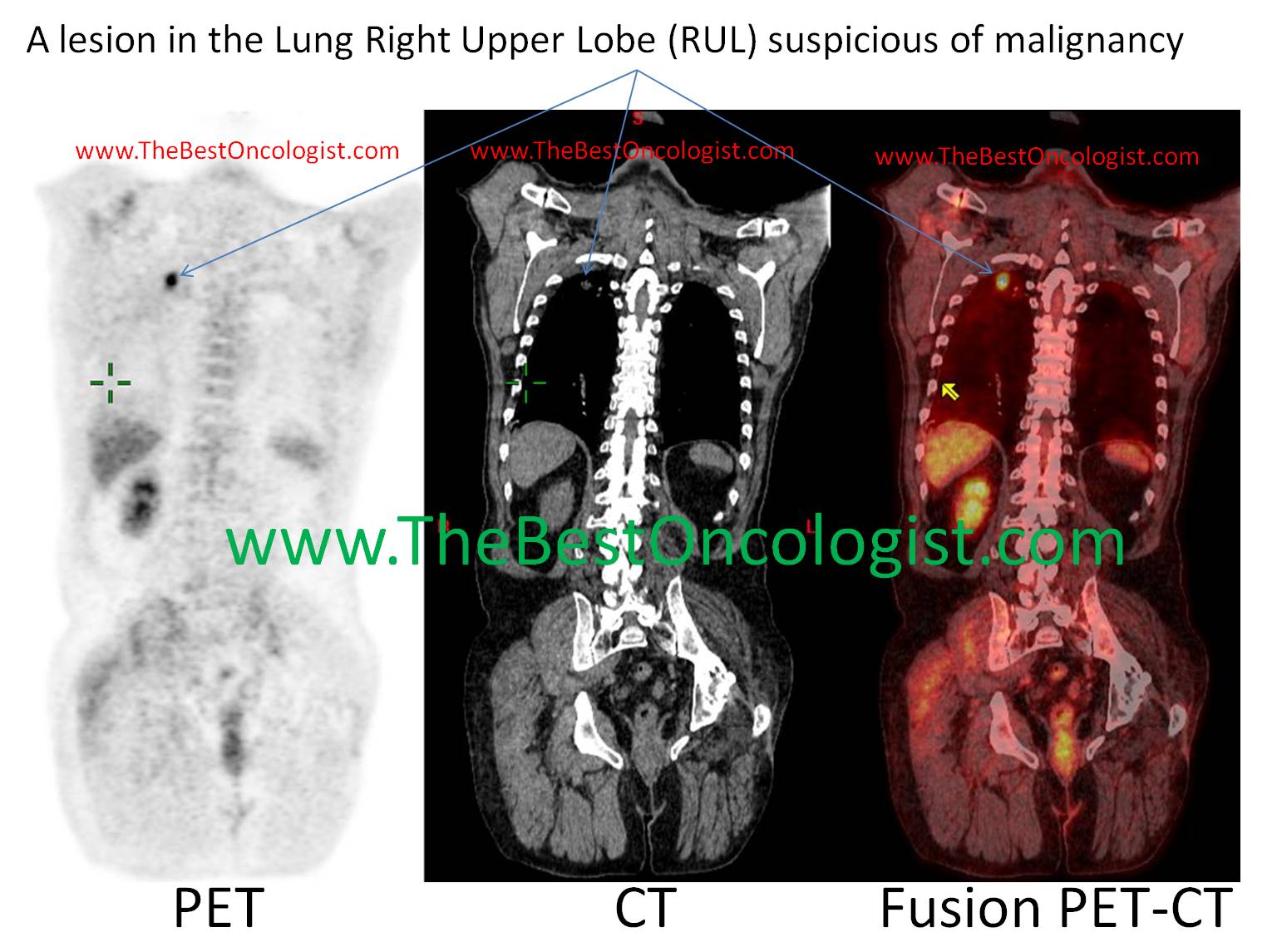
A lesion in the
Lung Right Upper Lobe (RUL) suspicious of
malignancy
5.
Pathological evaluation makes the final
diagnosis. Tissue biopsy should be obtained. Several methods are
available to get material for pathologic examination:
A.
Sputum cytology: the sputum is examined
under microscope for the presence of malignant cells.
B.
Bronchoscopy: in this method direct
inspection of the bronchial tree is performed, and biopsy is taken
directly from the tumor. Alternatively, the bronchi are washed with
saline, which is recollected and tested for malignant cells
(cytology).
C.
Fine needle aspiration (FNA): the lesion is
approached via a fine needle under the guidance of a radiological
facility (ultrasound or CT). The aspirated material is tested for
malignancy under microscope.
D.
Open biopsy: this is performed in operation
setting. This approach is usually good for lesions that can’t be
approached by FNA, and for localized tumors that may be totally
removed by surgery (tumors localized to single lobe, with no
metastasis).
|